
Image analysisbuilt forscientists.
Stand on the shoulders of your community. Streamline microscopy image analysis and cell segmentation with scientist-trained algorithms.
Built by people trained at


Take the guesswork out of your microscopy workflow
Manual analysis is less reliable than most researchers realize. Get instantaneous, validated, reproducible results across a variety of assays, from cell counts to IHC and ICC image quantification.
We got tired of unintuitive tools and incomplete workarounds.
So we built one platform that does it all.
HalfPage was built by scientists, for scientists. We responded to every frustration by building a feature for you.
Simple by design
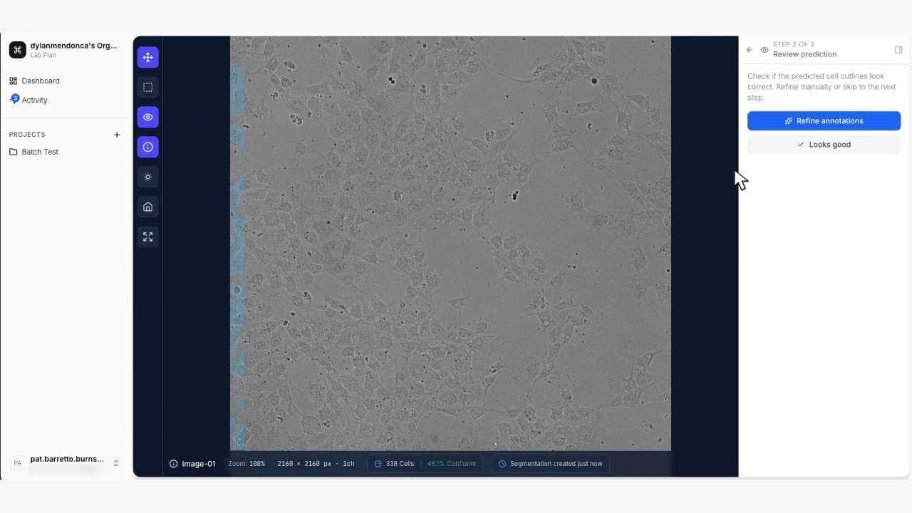
Existing tools demand powerful workstations, expensive licenses, and coding skills most researchers don’t have. We built HalfPage to run entirely in your browser: no installation, no hardware requirements, no programming needed. Point at a cell. That’s the whole workflow.
Results in seconds
Setting up a new analysis pipeline for every experiment wastes weeks of research time. HalfPage gives you access to community-trained models ready to use instantly: no learning curve, no trial and error. Quantified results in less time than it takes to make coffee.
Community-tested accuracy
Results that can’t be reproduced across labs aren’t results at all. Every HalfPage model is trained and stress-tested on diverse datasets contributed by researchers worldwide. The more you use the algorithm, the better it gets.
Built for your whole lab, and your collaborators too
Share your workflow with your colleagues whenever you're ready. Confirm your findings across the lab, inside the institution, around the world. Build the method everyone trusts and relies on.
Built for the images your lab already captures
HalfPage helps scientists analyze microscopy images, including fluorescence and brightfield captures from immunohistochemistry (IHC) and immunocytochemistry (ICC) experiments, with community-trained cell segmentation.
Microscopy image analysis
Upload brightfield or fluorescence microscopy images and run scientist-trained segmentation models in your browser, with no workstation setup or coding required.
Cell segmentation & quantification
Segment cells and nuclei, then measure confluence, counts, marker colocalization, viability, and related assay readouts with reproducible results.
IHC & ICC imaging workflows
After immunohistochemistry or immunocytochemistry staining and imaging, use HalfPage to segment cells and quantify what your stains show.
Microscopy analysis issues
While manual counting can be slow or inconsistent across the lab, our standardized models give you validated, shareable quantification instead of one-off pipelines.
Analyze. Adjust. Export.
Use scientist-trained models to analyze your data. Adjust it for your biology. Get your results. No coding, no setup, no expensive hardware or training time required.
Simple, Transparent Pricing
Free Plan
IndividualFor individual researchers
Pro Plan
Most UsedFor individual power users
Lab Plan
EnterpriseFor biology labs
About HalfPage


HalfPage was born from our frustration with the current state of scientific image analysis. As researchers, we experienced firsthand the challenges of working with imaging data. We are a team of cell biologists, computer engineers, and software developers focused on making your research faster, easier, and more reproducible.
We built HalfPage to solve these problems, creating a platform that's powerful yet accessible, with validated models that produce reliable results regardless of your technical expertise.
Ready to transform
your research pipeline?
Join scientists who are accelerating their research with scientist-trained algorithms. No coding required.
Get in Touch